Out of nowhere, a woman’s right arm and leg started shaking uncontrollably. In the moments leading up to the experience, she seemed well; she had no headache, confusion, or weakness. But for about a minute, half of her body juddered.
The woman, a rice farmer in the Philippines, headed to the emergency department. There, doctors gave her a physical exam and found nothing out of the ordinary. Then they took a look at her brain. Magnetic resonance imaging revealed a bright cluster of nodules surrounded by fluid in her left frontal lobe. The doctors had an immediate guess as to what they were: worm eggs.
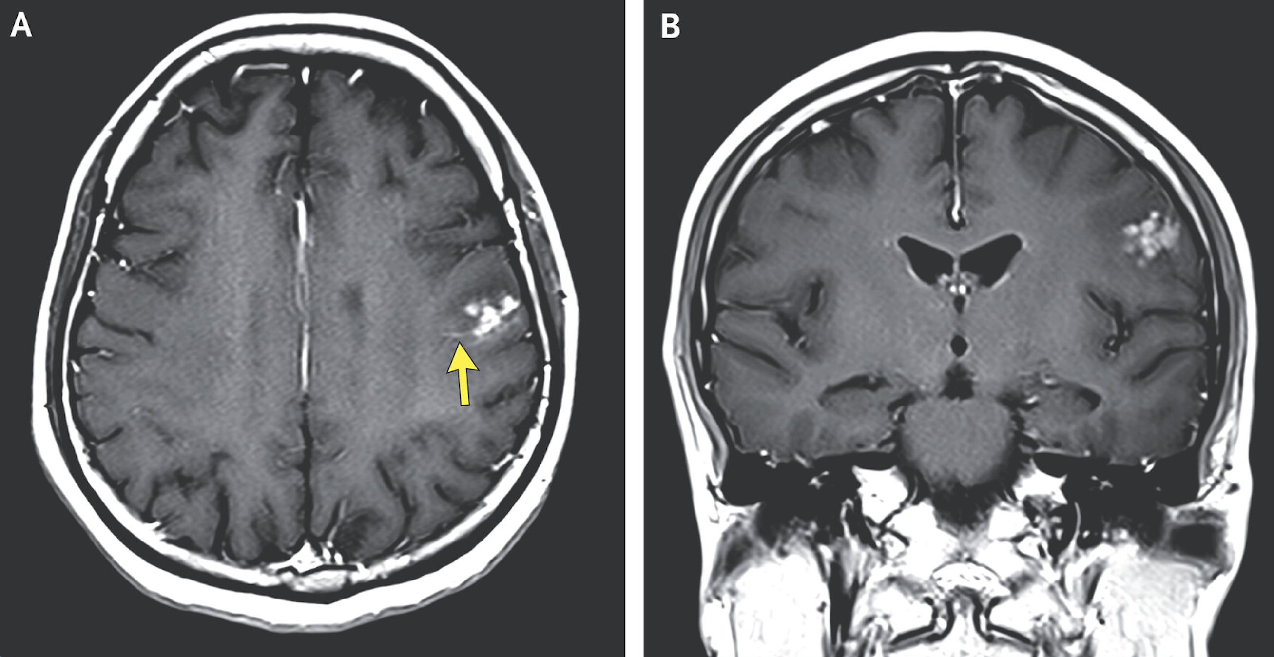
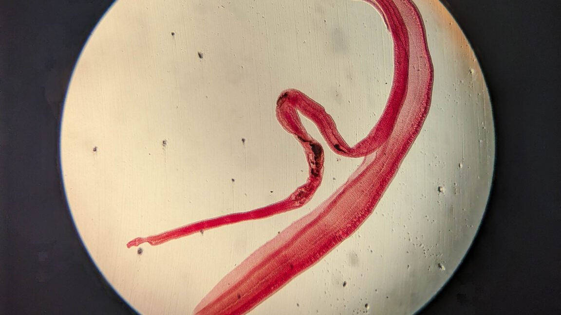
In a case report published in the latest issue of the New England Journal of Medicine, doctors write that they suspected the woman had neuroschistosomiasis, an uncommon complication of an infection with a Schistosoma parasitic worm, aka a blood fluke. In the Philippines, the most common fluke is S. japonicum. It’s also found in China and part of Indonesia; despite its name, it has been eliminated from Japan.
An unfortunate fluke
S. japonicum, like its relatives, has a diabolical life cycle, which starts with its eggs being shed in the urine or feces of various animals, including humans. When conditions are right, the eggs hatch into a free-swimming larval form. These seek out specific types of aquatic snails, which they bore into and inhabit to go through two life stages. Late-stage larvae then burst out, grow a forked tail, and swim to find an animal host. These tailed larvae, called cercariae, have the ability to tunnel through intact skin and take a trip through the vascular system—visiting the lungs, heart, and then liver, where they develop into adults.
The adult male and female worms live paired together with the slender female worm tucked into a groove within the thicker male. Adult worms typically move to large blood vessels in the abdomen connecting the liver to the intestines. From there, the cuddly couples spew out eggs that make their way into feces and urine.
Adult worms often go undetected in blood vessels, producing no noticeable symptoms, allowing the infection to become chronic. It’s the eggs that typically cause problems and give the worms’ presence away. They can spur inflammation, fibrosis, and organ-specific complications.
Ectopic eggs
On rare occasions, S. japonicum eggs reach the central nervous system and cause problems. It’s not entirely clear how this happens; medical experts speculate that it may happen when either the adults or eggs get diverted into an unusual place, possibly due to a heavy infestation or vascular damage. However it happens, when eggs get lodged in the brain, they’re often linked to seizures or specific neurological problems.
The possibility of S. japonicum eggs in the woman’s brain seemed like it could explain her abrupt seizure, so her doctors began diagnostic tests. There is no single go-to test for schistosomiasis, and doctors often do various tests to confirm an infection. The woman’s doctors first turned to an old-school lab test called the circumoval precipitin test, which was developed in the 1950s. In this test, doctors obtain a reference sample of S. japonicum eggs and flood them with a patient’s serum. If the patient has an infection, their serum will contain antibodies that can target the egg. In this event, the antibodies can grab onto molecules dangling off the outside of the egg as well as molecules secreted by the egg. In the test, this leads to a tail-like structure of bound antibodies forming on the outside of the egg.
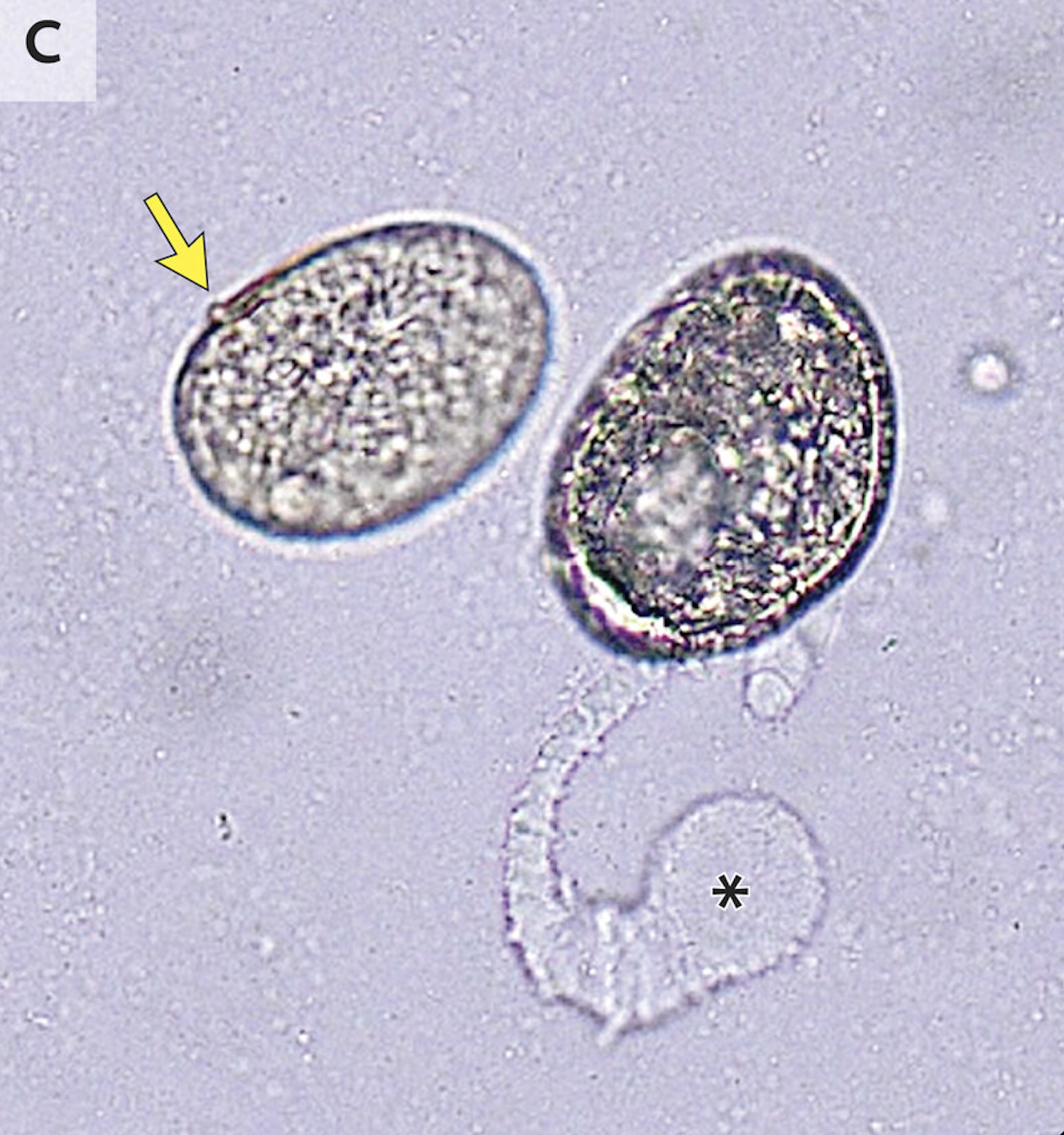
This test doesn’t always work, but in the woman’s case, it produced a clear result: test eggs grew weird tails, indicating an S. japonicum infection. Another test also found a lot of S. japonicum eggs in the woman’s stool: 190 eggs per gram of feces, to be precise. Together with the MRI result suggesting the presence of eggs, they diagnosed her with cerebral schistosomiasis that caused a focal seizure.
The doctors prescribed her an anti-parasitic drug, an anti-inflammatory drug, and an anti-seizure medication. Three months later, the woman told doctors she had no additional seizures. A follow-up MRI showed the worm eggs were gone.