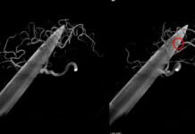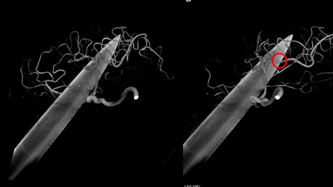
Despite the ghastly injury, the man was alive and conscious when he arrived. He could still talk and move all four limbs. His doctors described him as being in a state of agitation. On the Glasgow Coma Scale, he scored a 12 (E3 V4 M5), indicating a “moderate” brain injury, just one point above “mild.” He could open his left eye when told, but otherwise kept it closed (E3 score); he could talk and answer questions, but was confused (V4); and he intentionally moved away when pressure was applied to his body (M5).
In a report published Friday in BMJ Case Report, his doctors said they were concerned that in his confused, agitated state he may try to pull the knife out on his own. So, they swiftly sedated and intubated him. They put him on prophylactic treatments for bacterial infections, including tetanus. Then they used images to see what was going on in his head.
“Pull-and-see”
The blade was entirely through the right eye socket and eyeball. It traversed the left frontal and temporal lobes of his brain, passing through in front of—and without apparently injuring—the corpus callosum, which connects the two sides of the brain and is full of nerve fibers. The blade also missed the left internal capsule, which holds another bundle of nerve fibers.
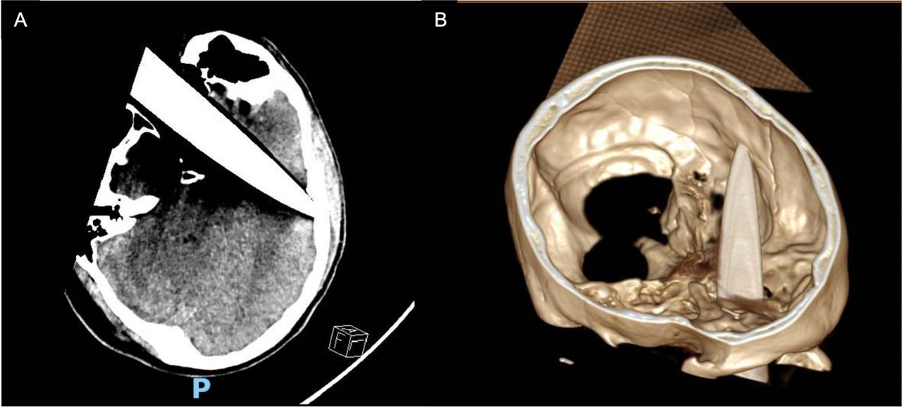
In terms of damage to major blood vessels, doctors found that the blade was close to the left internal carotid artery, a major blood vessel, but didn’t seem to have damaged it. Doctors did find, however, that a branch of the middle cerebral artery looked to be indented by the knife.
After consulting with various medical specialties, the man’s doctors moved to emergency neurosurgery. They opted to remove part of his skull to directly see into the brain, but used a conservative “pull-and-see” method to slide the blade out rather than invasively excavate it. After it was out, the doctors saw no signs of significant hemorrhaging. The brain wound was cleaned and drained before surgeons patched up his skull. His right eye, however, couldn’t be saved. Given its extensive damage, it was removed.
The man then moved to intensive care, where he recovered rapidly. A day after surgery, doctors were able to remove his breathing tube. He developed some minor right-side weakness, but it resolved two days later. Scans of his brain showed steady improvement—a little bleeding and swelling, and there was some air in his brain tissue from where the knife had been. But otherwise, it appeared to be healing. He developed no infections and had no seizures.
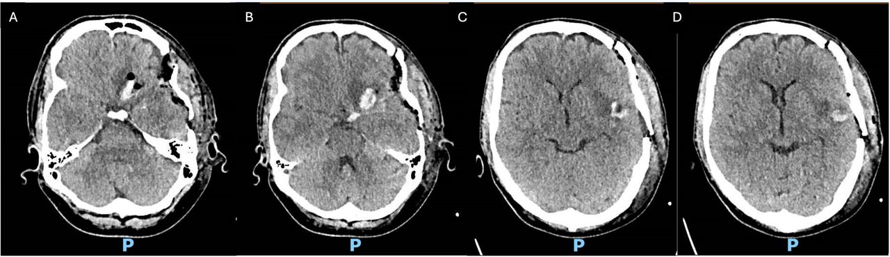
Despite the “dramatic intracranial trajectory,” his language function was preserved, and he had no loss of movement or coordination, they reported. He was discharged from the hospital and at follow-up appointments remained “neurologically well,” with ongoing eye and mental health care.
Lucky recovery
As for how the injury happened, his doctors still don’t know. “The mechanism is unclear,” they wrote. However, they noted he had a history of depression.
The doctors reflected on how lucky the man was to have survived and to have such a full recovery. Such injuries that go through the eye and the brain are rare. When they do occur, they’re usually from “high-velocity mechanisms” such as gunshots or other ballistic injuries. Knives, rods, and nails fit into the rarer “low-velocity mechanisms. “Such “low-velocity penetrating brain injuries carry substantial morbidity and mortality because of hemorrhage, vascular injury, abscess, meningitis, cerebrospinal fluid leak, seizures and neurological deficit,” they wrote.
But such fortunate recoveries aren’t unheard of; they cited reports of other remarkably unexpected recoveries from seemingly devastating brain injuries. That includes the famous case of Phineas Gage, a railroad foreman who survived an 1848 accident that sent a large iron rod directly through his head. The rod entered below his left cheekbone and exited the top of his skull. (Although he survived and recovered, Gage’s personality was said to have changed after the accident. The doctors in the knife-wound case did not mention any such personality changes.)
“Echoing classic and contemporary examples of severe frontal lobe penetration with unexpectedly preserved neurological function, including Phineas Gage and a reported nail-gun injury, this case highlights that outcome may depend less on external severity than on precise trajectory relative to critical vascular, cortical and subcortical structures,” the doctors concluded.
Source: Iwpost.com