A 63-year-old woman arrived at a hospital in pain. She had abruptly lost vision in her right eye. As doctors got to work, the series of findings that followed revealed not just bad news for her, but an alarming revelation of how infections can evolve within a patient.
The woman’s remarkable case was reported this week in the New England Journal of Medicine. According to her doctors, initial magnetic resonance imaging scans of her brain quickly identified the key problem. The imaging indicated inflammation in her right eye, but also picked up lesions in her brain. Specifically, the imaging suggested problems in her parietal lobe, which is a hub for processing sensory information.
The doctors suspected she had a brain abscess. Laboratory results of her blood and urine samples suggested an infection as well as kidney problems. Doctors decided to open up her skull for a brain biopsy. Inside, the surgical team was met with pus. Meanwhile, doctors worked to identify the bacteria found in two urine samples.
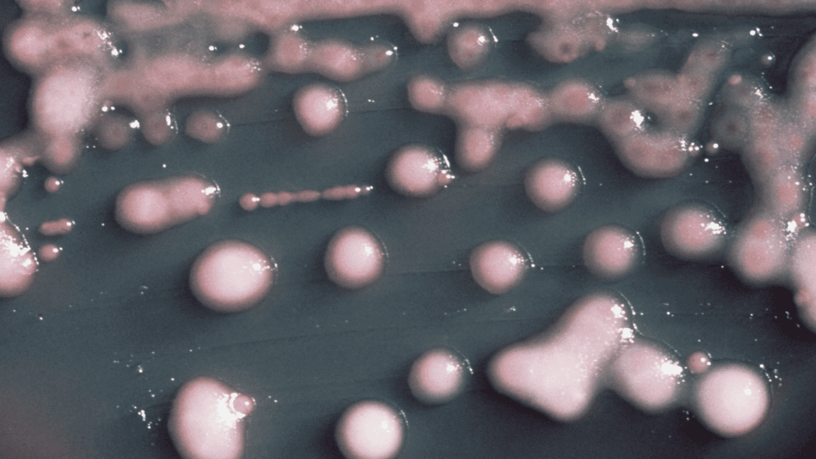
In all, the two urine samples and the brain sample all grew the same bacteria: Klebsiella pneumoniae. This is a bacterium well known to doctors. It often dwells in the gastrointestinal tract, but can lurk in healthcare settings and spark infections, particularly pneumonia and urinary tract infections. It’s also known for having a variant that can go metastatic, traveling via the bloodstream to invade other organs, particularly the liver, but also the lungs, eyes, and brain.
In a standard case report, this might be the end of the story. Doctors would diagnose a disseminated K. pneumoniae infection and treat with antibiotics. But there was more.
Goopiness
Doctors and researchers brought in to help with the case noticed that although the three samples all grew K. pneumoniae, the bacteria were different. Bacteria in the first urine sample looked like normal K. pneumoniae. But the bacteria from the second urine sample and the brain were gloopy and sticky. Under an electron microscope, the doctors could see that the K. pneumoniae in these two samples had thick capsules—they were seemingly encased in goop.
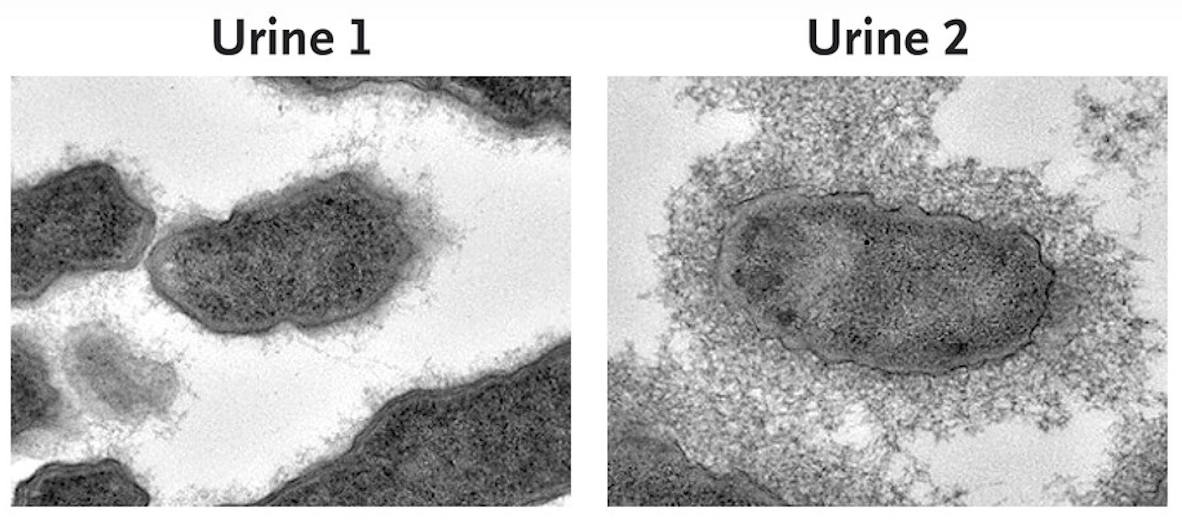
Further tests showed that the thick sticky coating made these bacteria more virulent than the normal-looking bacteria from the first urine sample. With the coating, protective immune cells called macrophages couldn’t gobble up the bacteria as they could with the normal K. pneumoniae. In mice, the snotty bacteria were lethal, while the normal one wasn’t. In all, the two sticky samples met the definition of hypervirulent K. pneumoniae—the kind known for going metastatic.
But being hypervirulent wasn’t all fun and brain-eating games for these bacteria; it had drawbacks, too, the researchers found. The hypervirulent bacteria couldn’t grow nearly as quickly. They also couldn’t infect cells in the bladder as well.
Genome sequencing of the three bacterial samples showed that they were all highly related. And, interestingly, they all lacked the hallmark genes associated with hypervirulence in K. pneumoniae. The bacteria in the two urine samples differed by 66 small mutations, while the two hypervirulent bacterial samples differed by just four small mutations. These two hypervirulent bacterial samples had key mutations in genes that produce the bacterium’s capsule, explaining their gloopiness. (The researchers confirmed this by genetically engineering normal K. pneumoniae to contain these specific mutations, which reproduced the goopiness.)
With the genome sequencing, the researchers estimated that the bacteria in the two urine samples diverged from a common ancestor one to two years before the woman’s current state. Then, more recently, the hypervirulent urine bacteria disseminated, spreading to the woman’s brain.
The woman’s doctors noted that two years before her brain abscess, she was hospitalized with a urinary tract infection.
Heterovirulence
Bacterial evolution within a single patient is not particularly new. Infecting bacteria often adapt to have a fitness advantage in their victim. But in most cases, the variant with the biggest edge overtakes the population. In less common cases, the population splits. This has been seen when bacteria are trying to survive antibiotic treatments. In this case, some variants become more resistant to the treatment, but the resistance comes at a cost—slower growth or less virulence, for example. This keeps them from dominating. Instead, they remain as a subpopulation. This scenario is called ‘heteroresistance,’ which is linked to treatment failure.
In the woman’s case, all three isolates were susceptible to antibiotics—some good news. She was able to be treated with antibiotics to clear the infection. Further, most patients with K. pneumoniae brain abscesses recover, though there were no follow-up details of how her neurologic or vision recovery went.
While her infection didn’t have heteroresistance, it had something new: heterovirulence. The hypervirulent subpopulation had a fitness disadvantage in the urinary tract—the origin of the infection. But it had an advantage for dissemination and virulence, spreading to the brain and eye.
Overall, the researchers and doctors report that “this case shows the ability of K. pneumoniae to disseminate within a patient from a clinical (urinary) reservoir to distant body sites and highlights the potential for the emergence.”