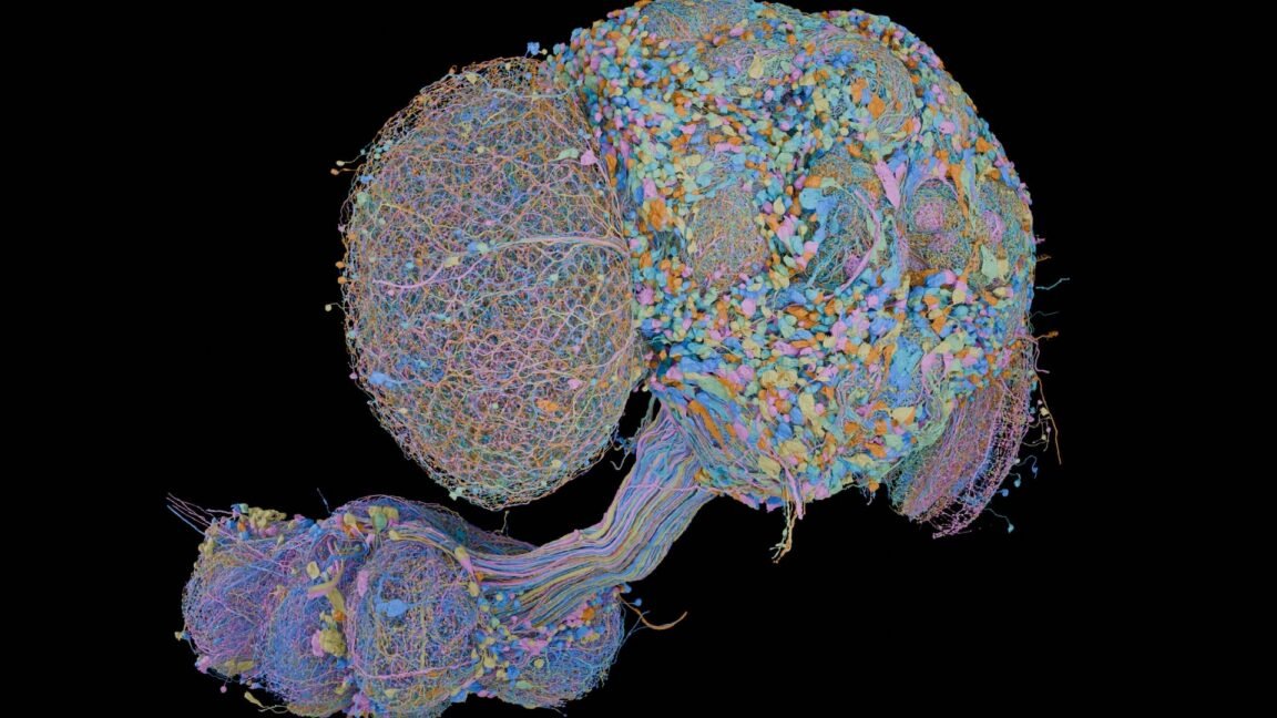
On Friday, researchers announced the completion of a map of every neuron in the brain of a male fruit fly. The “connectome” provides a tool that can accelerate neurobiology research. But it also provides an opportunity to do some science on its own, as the connectome of a female Drosophila had been completed earlier this year. The work also provided the team behind it the opportunity to refine tools that are likely to be applied to ever-more complex nervous systems, including (potentially) those of vertebrates.
The new work involved a collaboration between biologists at the Howard Hughes Medical Institute’s Janelia Research Campus and computer scientists at Google—both acknowledge that neither could have done the project without the other. Preparation of an entire brain for imaging at the necessary resolution requires a distinct set of skills, as does interpreting what those images indicate. But building a complete picture of the hundreds of millions of synapses in a brain as small as the fruit fly’s is a task that can’t be achieved by humans in a manageable amount of time.
The people behind the effort expect that in the long term, the effort will be worth it, as the connectome could give neurobiologists a valuable tool for understanding how the brain works.
Establishing a connectome
Our interactions with the world begin with sensory input—the neurons that register sound, light, touch, and more. From there, most brain activity involves neurons communicating with each other. This communication transforms the inputs into signals the rest of the brain can interpret, routes information to relevant processing centers, and often produces some kind of output, from forming a memory to moving a muscle.
All that processing is dictated by which neurons have connections to others. For example, the visual system does some basic recognition of its own before passing the results to the brain’s visual processing centers. If those centers detect something like text, they can use connections to the language centers to interpret it, and so on.
To understand how a brain works, then, we need a catalog of the connections in the brain, since those dictate how information flows through its various systems. That catalog is a connectome.
In practical terms, a connectome is the list of every neuron in a brain, including its location in three-dimensional space, and the connections (termed synapses) it forms with other neurons. That’s more complicated than it may sound. Each neuron can form multiple, branched processes called axons, allowing it to form hundreds of connections to other neurons. So while the nervous system of the fruit fly consists of only roughly 150,000 neurons, and the brain contains only a fraction of those, the new work discovered over 300 million synaptic connections in the fly brain.
So how do you go about mapping something like that? Gerry Rubin, a senior group leader at the Janelia Research Campus and one of the senior authors on the new paper, described how things have changed considerably based on the complexity of the system. “I was a graduate student at the [UK’s Laboratory of Molecular Biology]… and when I got there in 71, they already bought this giant computer, and they had the idea that they were going to use machine vision and computers to assemble the C. elegans connectome,” Rubin said.
C. elegans is a small, transparent worm with just over 300 neurons and would seem to be a tractable system. “It took them about two years to realize that the computers were nowhere near powerful enough,” Rubin said, “and so they went with printing everything out on photographic prints and colored magic markers and circling neurons and tracing it by hand.”
That level of attention would simply not work for something as complex as the fruit fly, which is also very much not transparent, making its nerves difficult to image. Fortunately, computers have advanced considerably, as have the algorithms we’re able to run on them.
For the new work, the biologists took a dissected fruit fly brain (along with part of its ventral nerve cord) and cut it into a huge series of evenly spaced slices. By knowing the exact order of the slices, the researchers were able to maintain the three-dimensional architecture of the brain even while converting it into a series of roughly two-dimensional objects that could be imaged using electron microscopy, provided the resolution needed to identify small cellular structures. From there, computers become essential to processing the images.
Putting AI to work
Michal Januszewski, a staff scientist at Google Research, told Ars that the first step is to use a form of generative AI to ensure that the areas at the site of each slice are linked up properly. No matter how carefully you slice, there will be some distortions and a bit of material lost when you make a cut.
“When you take those blocks and you stitch them back together computationally, there’s a little bit of a gap in between them so the tissue doesn’t completely smoothen,” Januszewski said. “We use [a generative] model to make the tissue look as if the seams were not there, and that then makes all the downstream processing easier because you can basically ignore the problem to a large degree.”
A separate model acts by filling in the spaces defined by cellular membranes, allowing the system to track individual cells across 3D space. “It is different in a number of ways from what people commonly think when they talk about AI,” Januszewski told Ars. “One is that it is actually a recurrent process, so it literally moves through space as it makes the outline of the neurons, and it is a visual model, so it converts voxels out of the images from the microscope, [converting them] into a 3D presentation of the neurons.”
Still, other models are used to recognize synapses and classify the type of synapse once it’s spotted. These models can be fine-tuned using different levels of sensitivity, or “greediness”—basically, adjusting the probability that they’ll call something a synapse. And here, feedback from human proofreaders plays a major role.
“This has to be tuned by going back and forth between the proofreaders and Mikhail to say, ‘Oh, give us a version where you were less greedy because it’s harder to disassemble than it is to assemble,’” Rubin said. “So it’s an iterative process between the humans giving feedback and the algorithms getting tuned.”
All of this took roughly four years to go from an intact fly brain to the complete connectome. But Rubin said the techniques the team developed along the way, along with the growing sophistication of the software, will hopefully be critical as connectomics work moves up the complexity scale.
“Our view is we did Drosophila with a team of 50 people,” Rubin said. “The hope is, by the time someone does a mouse, they’ll also need a team of 50 people, even though there are a thousand times more neurons in there. The people will never go away, but the people will not need to scale with the number of neurons, which would be economically not feasible.”
Managing this complexity is also what drew Google to the challenge. “The reason I think Google and we were interested in this is because this is this type of grand challenge that just cannot be done in any other way,” Januszewski said. “We knew we need AI for this. This cannot be solved by having more humans or by any other technology. And it’s important.”
Sex on the brain
Due to the extensive history of research on Drosophila, we already knew a great deal about the fly nervous system, including the functional regions of the fly brain and an assortment of individual neurons that had been identified by a combination of function and/or gene activity. But it was at best a partial picture, one that we can use the connectome to fill out in more detail. Combined with the completion of the connectome of a female fly that was completed by university-based researchers earlier this year, we’re able to understand a bit more about how sex determination feeds into specific brain differences.
And as with so many things in biology, the basic numbers look simple but the details are fairly complex. The researchers identified 289 male-specific neurons, 71 female-specific ones, and 138 that were present in both sexes but formed a different shape and connections in males and females. Their relationship to the genes mentioned above was not always direct. Ninety percent of the male-specific neurons were making dsx and fru, but that leaves 10 percent that weren’t. Rubin suggested these had likely picked up a sex-specific identity by interactions with those that were.
That may also be true for the neurons that were present in both sexes but which had different shapes and connections. Nearly 40 percent of these neurons did not have active dsx or fru genes. And about 7 percent of the neurons that were identical in the two sexes did have dsx and fru activity, suggesting that they may have some other difference in neural activity despite forming a similar shape and connections.
There were also some oddities. For example, two neuron types that control the female’s physical response to mating also showed up in males and had the same basic shape, even though the tissue that they indirectly control doesn’t even exist in males. The neurons instead differed in the connections they made in males and females.
The research team also identified a general pattern in where these sex-specific neurons act in neural pathways: They tended to be removed from immediate sensory processing or motor control, instead acting in concert with neurons engaged in higher-level neural processing. There are a handful of exceptions, such as sensory neurons that register the presence of sex-specific pheromones. But for the most part, the neurons appear to act after basic sensory processing is complete, potentially bridging that processing with relevant behaviors.
In keeping with this, the sex-specific neurons tend to cluster together, suggesting that they may act together to enhance the intensity of the signals they convey. As the paper puts it, the location and connections of the sex-specific neurons “suggests a hierarchy in which sex differences primarily modify integrative and decision-making areas while sensory detection and the highly tuned motor interface remain more constant.”
What’s next?
In some ways, the completion of the male and female Drosophila connectomes will be a bit like the completion of the fly genome: It will accelerate a lot of work that was already underway. If a study identifies an interesting neuron, researchers can simply open a browser and determine which other neurons and brain structures it connects to. In difficult-to-quantify ways, that should lead to more informed hypotheses while saving researchers the work needed to understand the neural connections.
Rubin also suggested it’s having a big impact on theoretical neuroscience. “Before this, most neuro theorists were very much like, ‘How could a brain work?’” he said. “And they didn’t have a constraint. Once they had the connectome, they could say, ‘The brain does this and here’s the wiring diagram. How can this wiring diagram allow this function?’ So this has been a major. I’d say this is the biggest change in having the connectome.”
Other work will depend on whether completing connectomes follows the trajectory seen in genomics, where the cost drops precipitously as techniques are refined and further automated. One of this work’s most intriguing findings is that a specific neuron seen in the female connectome was absent from one of the two hemispheres of the male fly, presumably due to a developmental glitch. With only two connectomes complete, it’s impossible to get a good sense of how common this sort of variability is. The same applies to subtler differences in the trajectories taken and connections made by individual neurons.
To say anything meaningful about this kind of variability with statistical confidence, we’ll need dozens of examples. That means we’ll also need to make the process much faster than the four years it took to go from dissecting a fly brain to the final connectome.
Cell, 2026. DOI: 10.1016/j.cell.2026.08.015 (About DOIs).